A normal reading-vision test or a single eye-pressure result cannot provide a complete glaucoma assessment. You will learn when to book screening, which tests to request, how to prepare, and which symptoms require emergency care rather than a routine appointment.
Key takeaways
- Book screening after 40 even when your vision seems normal.
- Seek urgent care for sudden eye pain, halos, nausea or blurred vision.
- A complete assessment includes pressure, optic nerve and visual field checks.
- Follow-up depends on your results, eye structure and glaucoma risk factors.
When should you be screened, and when is it an emergency?
Age 40 increases your need for glaucoma attention, but it does not create one testing interval for everyone. Early glaucoma is often symptom-free, so good reading vision does not exclude it.
If your first examination is normal and you have no major risk factors, ask the examining clinician when to repeat it; timing depends on your age, optic-nerve appearance, eye pressure, corneal thickness and medical history.
Request earlier or more frequent review if you have:
- A parent, sibling or child with glaucoma
- Diabetes or high blood pressure
- High myopia (short-sightedness)
- Steroid use, including creams, inhalers, tablets, injections or eye drops
- A previous eye injury or surgery
- Thin corneas
- Advancing age or a previously raised pressure or suspicious optic nerve
Sudden severe eye pain, a red eye, blurred vision or halos around lights, headache, nausea or vomiting, or sudden vision loss can signal acute angle closure. Seek emergency eye assessment immediately; do not wait for a routine glaucoma checkup in Mumbai, because pressure-related damage can progress rapidly.
For glaucoma screening for over forty in Mumbai, remember that population-wide screening evidence is not definitive. A useful appointment balances your risk factors, examination findings, false-positive results and the ability to provide follow-up, rather than promising that one pressure reading prevents blindness.
Which tests belong in a complete glaucoma assessment?
A complete glaucoma test for adults over 40 in Mumbai combines pressure, anatomy and function. Good visual acuity only shows how clearly you read; it does not rule out early optic-nerve damage or visual-field loss.
- Visual acuity checks reading and distance vision.
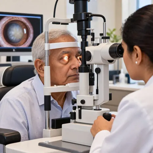
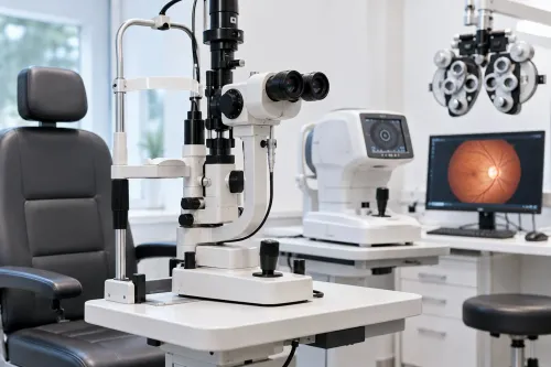
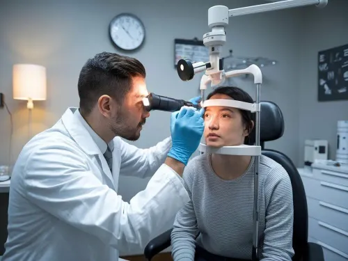
- Slit-lamp examination inspects the cornea, lens and front of the eye.
- Calibrated tonometry measures intraocular pressure.
- Optic-nerve examination looks for cupping, asymmetry and damage.
- Gonioscopy uses a special lens to determine whether the drainage angle is open, narrow or closed. It cannot be replaced by an eye pressure test in Mumbai.
- Central-corneal-thickness measurement, or pachymetry, records corneal thickness for pressure interpretation and risk assessment.
- Automated visual-field testing, or perimetry, measures functional loss.
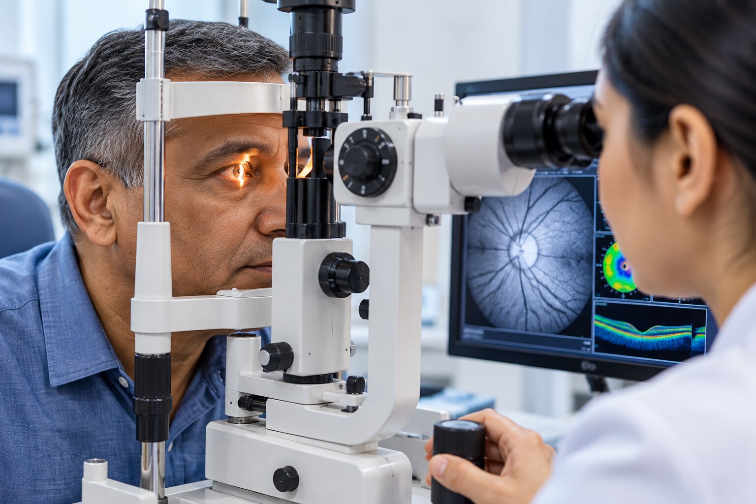
- OCT, when indicated, images the retinal nerve-fibre layer and ganglion-cell layers as structural evidence.
OCT scans can contain segmentation or image-quality artefacts, while visual fields are affected by fatigue, poor fixation, uncorrected refractive error and learning effects. A first abnormal scan or field may need confirmation.
| Test option | What it contributes | What it cannot establish |
|---|---|---|
| Pressure-only kiosk | An intraocular-pressure reading | Whether the angle, nerve or field is abnormal |
| Automated eye photograph | A captured image of the eye or optic disc | Reliable functional loss or a complete diagnosis |
| Full assessment | Pressure, angle, nerve, cornea, field and indicated OCT findings | A guarantee that future disease cannot develop |
What happens during the pressure test, and how should you prepare?
An eye pressure test in Mumbai is tonometry: it measures intraocular pressure, the pressure inside your eye, not your vision or optic-nerve damage. It is one part of a glaucoma checkup in Mumbai, not a diagnosis by itself.
- For a non-contact test, keep your forehead and chin against the supports while a brief puff of air reaches the eye. The air-puff instrument does not touch you.
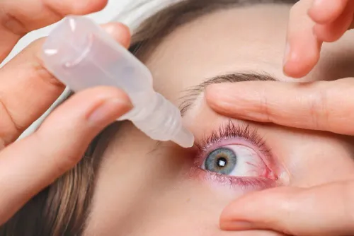
- For applanation tonometry, the clinician puts in anaesthetic eye drops, cleans the instrument, and briefly touches the eye’s surface with the measuring tip while you look steadily at a target.
- Tell the clinician if you wear contact lenses and ask whether you must remove them before testing. Bring previous pressure readings, visual-field printouts, OCT scans, eye-operation records and a complete medication list.
- Include steroid creams, inhalers, tablets, injections and eye drops. The route does not determine whether a medicine affects glaucoma risk or pressure interpretation.
- Ask what happens if the result is borderline or unexpectedly high. Eye pressure changes during the day and between visits, so the reading may need repetition and interpretation alongside the optic nerve, drainage angle, corneal thickness and visual field.
Normal pressure does not rule out glaucoma: optic-nerve damage can occur within the usual population range. A routine spectacle vision test measures reading clarity; a comprehensive eye examination is broader, while a glaucoma assessment specifically evaluates pressure, drainage angle, optic nerve, cornea and visual field.
How are open-angle risk, angle-closure risk and glaucoma suspects handled?
One pressure reading cannot establish glaucoma: diagnosis depends on whether pressure, the drainage angle, optic-nerve appearance, structural imaging and visual function agree over time. Open-angle assessment uses the optic nerve, pressure pattern, visual-field results and OCT, with gonioscopy confirming that the angle is open.
| Pattern | Main evidence | What changes |
|---|---|---|
| Open-angle risk | Optic nerve, pressure pattern, OCT and visual fields; open angle on gonioscopy | Compare findings over time and confirm doubtful results |
| Angle-closure risk | Crowded or closed angle on direct gonioscopy | Use a different urgency and treatment pathway |
| Glaucoma suspect | Raised pressure, suspicious nerve or thin cornea without confirmed field loss | Assess risk and repeat or monitor rather than label from one result |
Angle-closure risk cannot be assessed by tonometry alone. Sudden eye pain, redness, halos around lights, nausea or rapid visual change belongs in emergency care because a closed angle can threaten vision quickly.
Pachymetry records central corneal thickness. It affects how measured pressure is interpreted and contributes to risk assessment, but the clinic should record the actual thickness rather than apply a simplistic universal correction.
OCT and automated fields must be read with the clinical examination; segmentation errors, poor image quality, fatigue or poor fixation can make a scan or field unreliable.
When comparing a glaucoma specialist in Mumbai, ask whether the service performs or arranges:
- Gonioscopy and calibrated tonometry
- Pachymetry and optic-nerve examination
- OCT and automated visual fields
Dr Nusrat Bukhari's Eye Clinic is one local option to evaluate against these capabilities. This is the standard to compare, not a substitute for comparing the assessment offered.
Related service
Glaucoma Screening & Management Glaucoma Screening & Management Glaucoma is a silent, progressive eye condition that can cause irreversible vision loss if not detected early. View service → |
What should you do after a normal or abnormal assessment?
A normal examination earns you a documented baseline and a repeat date, not a permanent all-clear. Ask for the recorded pressure, optic-nerve findings, angle status, corneal-thickness result, visual-field reliability and whether OCT was needed. Then obtain a specific review date based on your age and risk profile.
A normal pressure-only reading is not a normal glaucoma assessment.
| Result | What it means | Next step |
|---|---|---|
| Normal full assessment | No confirmed structural or functional damage today | Record the findings and follow the clinician’s stated repeat date |
| Raised pressure | Pressure needs interpretation alongside corneal thickness, angle and optic nerve | Ask whether to repeat measurements at another time of day |
| Suspicious optic nerve | The nerve appearance needs correlation with pressure, OCT and fields | Ask which finding requires confirmation |
| Abnormal field | The result may reflect glaucoma or testing difficulty | Ask whether fatigue, poor fixation, uncorrected refractive error or learning affected reliability |
Ask which result would change the follow-up plan. A reliable baseline allows future OCT scans and visual fields to be compared; one abnormal field is not definite progression if testing problems could explain it.
Clarify which symptoms require emergency care and which can wait for scheduled review. Sudden severe eye pain, a red eye, halos, nausea or vomiting, or rapid vision loss needs emergency assessment. Otherwise, keep the planned appointment.
Screening is a process, not a one-time guarantee. Whether you arrange glaucoma screening for over forty in Mumbai, a glaucoma checkup in Mumbai or review with a glaucoma specialist in Mumbai, choose documented follow-up over a clinic advertising only an eye-pressure check.
Frequently asked questions
When should adults over 40 have glaucoma screening in Mumbai?
Book a baseline assessment after age 40, then follow the interval recommended from your eye pressure, optic nerve appearance, visual field and risk factors. Do not wait for symptoms because early glaucoma often causes none.
When is glaucoma an emergency?
Seek urgent eye care for sudden severe eye pain, a red eye, blurred vision, coloured halos around lights, headache, nausea or vomiting. These symptoms can indicate acute angle-closure glaucoma.
Which tests are included in a complete glaucoma assessment?
A complete assessment commonly includes tonometry for eye pressure, optic nerve examination, gonioscopy to inspect the drainage angle, visual field testing and optic nerve imaging such as OCT when indicated.
How should I prepare for an eye pressure test?
Bring your glasses, medication list and previous eye reports. Tell the clinician about glaucoma in your family and any steroid use. Avoid tight clothing around your neck and follow instructions about contact lenses.
What happens after a normal or abnormal glaucoma assessment?
A normal result still needs follow-up at the interval your clinician sets. An abnormal result may lead to repeat measurements, additional imaging, visual field testing, treatment or referral to a glaucoma specialist.
Keywords